RMN Bucuresti

RMN Bucuresti

Medical Imaging

Located at Bulevardul Nicolae Grigorescu 41 in Sector 3, the clinic is strategically positioned for easy accessibility.

Contraindications for MRI:

Soft Tissue Imaging: MRI is particularly effective in imaging soft tissues and is invaluable in neurological, musculoskeletal, cardiovascular, and oncological imaging.

Types of MRI Examinations:

Contraindications: Patients with pacemakers, certain types of hearing aids, and certain metal implants may not be suitable for MRI due to the strong magnetic field. MRI is generally avoided during the first trimester of pregnancy.

Patient Care and Comfort:

Info

Imaging Techniques

Imaging Techniques

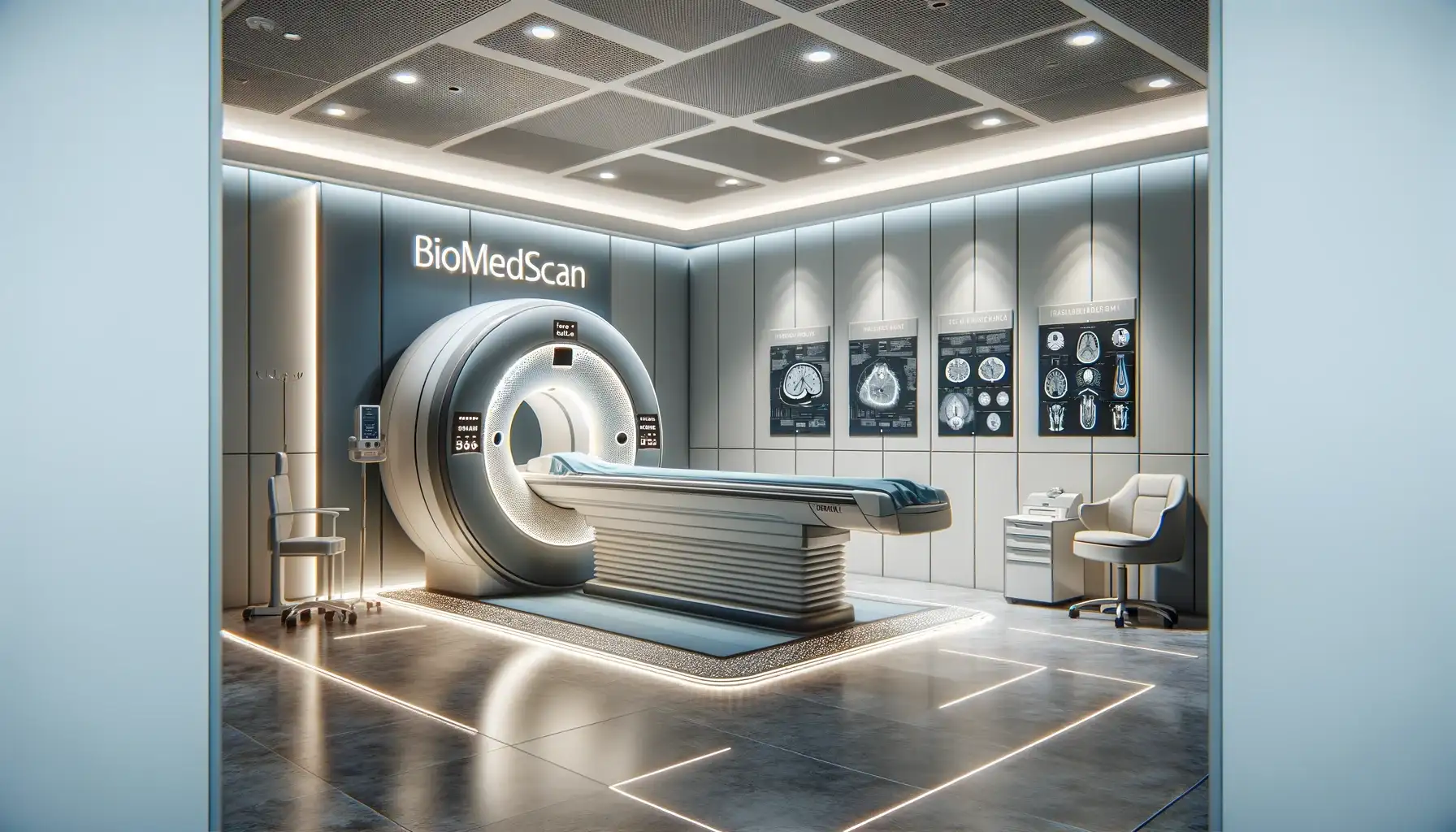
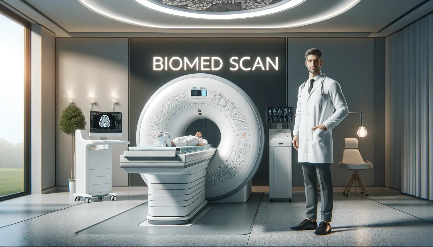
Biomed Scan Clinic, a prominent MRI center in Bucharest, has been providing exceptional imaging services for over nine years.

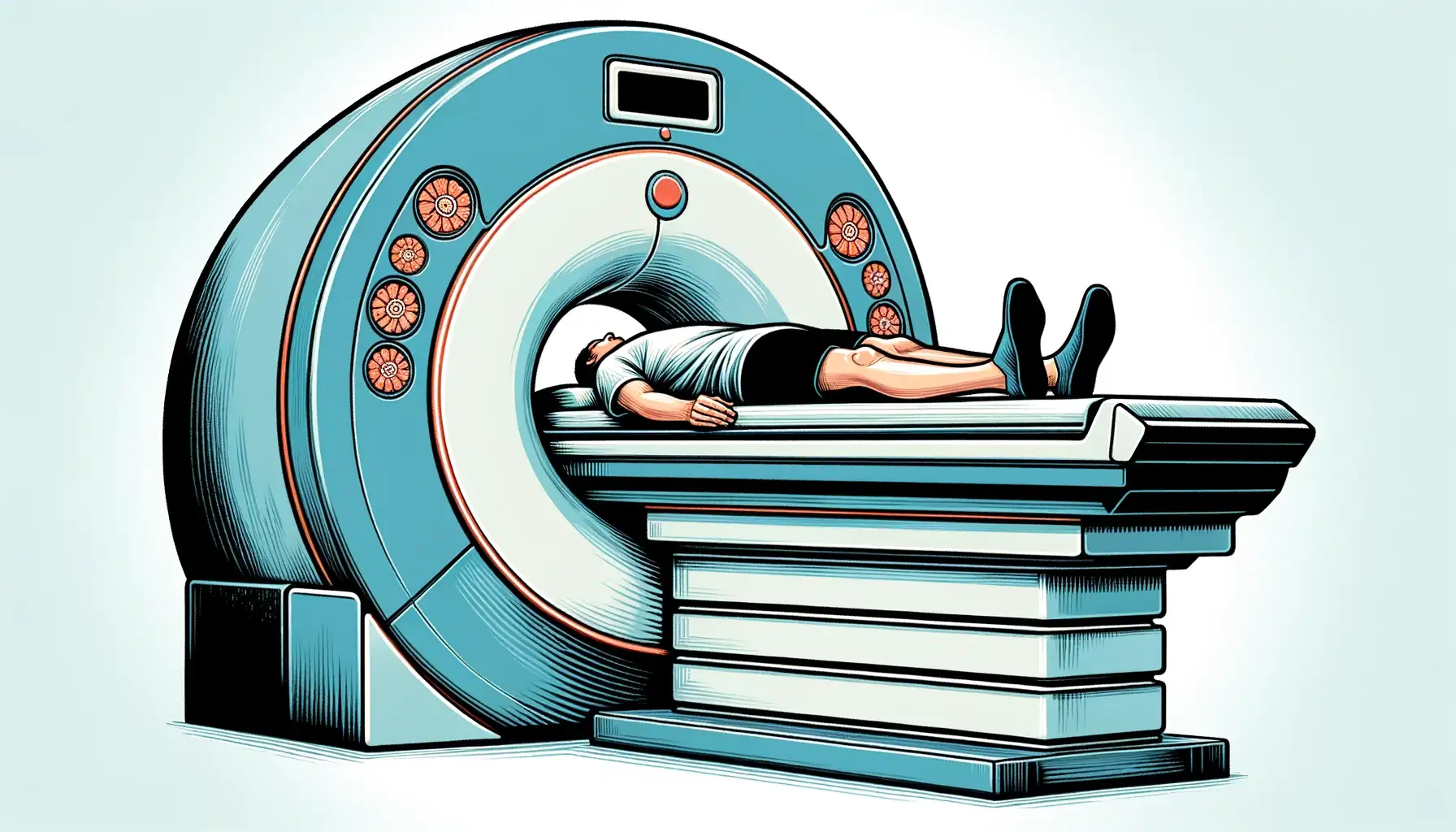
Preparation: Patients are required to remove all metal objects from their bodies, including hearing aids, dental plates, jewelry, watches, and hairpins, due to the strong magnetic field used in MRI.

Specializing in a broad spectrum of MRI-based imaging techniques, the clinic has served over 120,000 patients, showcasing a vast experience in various pathologies. The clinic's commitment to advanced imaging technology provides significant anatomical and physiological information, often complementing other diagnostic methods like CT, radiography, or ultrasound.

RMN Bucuresti

This location, known for its state-of-the-art medical facilities, has been home to Biomed Scan's journey in delivering top-notch diagnostic imaging services to its patients​​.

Biomed Scan Clinic, operating in Policlinica Titan in Bucharest for over 9 years, has firmly established itself as a leader in MRI (Magnetic Resonance Imaging) diagnostics.

Contrast Agents and Administration: The clinic uses gadolinium-based contrast agents like Dotarem, Gadovist, and Multihance, depending on the specific requirements of the examination. These agents enhance the clarity and detail of MRI images, aiding in more precise diagnoses.

Magnetic Resonance Imaging

Radiology

Robes and Privacy: Patients are provided with robes and are required to change clothes depending on the area being examined.

Safety Measures and Protocols:

Diagnostic Capabilities:

Radiology
Neuroimaging

Isolation and Supervision: During the MRI scan, patients are isolated in the examination room but are continuously monitored through a transparent window.

Documentation: Patients are advised to bring all relevant medical documents related to the area being scanned.

Metal Implants: Certain metal implants, such as some types of surgical clips, may pose a risk in the MRI environment.

Radiographer

Comparison with Other Imaging Modalities:

Specialized MRI Techniques:

During the Scan: Patients will hear various noises from the MRI machine and will be provided with earplugs to reduce noise. They are required to remain still throughout the scan to ensure image accuracy.

Radiographer

Frequently Asked Questions


  • MRI is a medical imaging technique that uses a magnetic field and computer-generated radio waves to create detailed images of the organs and tissues in the body. 
  • It's widely used in hospitals as a diagnostic tool, particularly for imaging the brain, spine, and joints.


  • In a brain scan, MRI uses a powerful magnetic field, radio waves, and a computer to produce detailed pictures of the brain's structure. 
  • It's an essential tool in neuroimaging, helping to diagnose and monitor brain disorders, injuries, and development.


  • T1 and T2 weighted images are types of MRI scans. T1 images are useful for assessing the anatomy of the brain, while T2 images help in identifying fluid and edema. 
  • These imaging techniques offer different views of tissues and are used based on the diagnostic requirement.


  • A contrast agent, often containing gadolinium, is used in some MRI exams to improve the clarity of the images. 
  • It helps to highlight certain structures or abnormalities more clearly, particularly in blood vessels and tumors.


  • A radiographer, or MRI technologist, is responsible for operating the MRI scanner, positioning the patient, and ensuring MRI safety protocols are followed. 
  • They also assist in optimizing image resolution and quality during the radiological examination.


  • Tesla is the unit of measurement for the strength of the magnetic field used in MRI scanners. 
  • Common clinical MRI scanners operate at 1.5 or 3 Tesla (3T MRI Scanner), but high-field MRI scanners can have strengths up to 7 Tesla or more, offering higher image resolution.


MRI sequences are sets of parameters that dictate how the MRI machine will operate to create images. Spin echo and gradient echo are types of sequences that manipulate the protons' behavior in the body, providing different types of image contrast and resolution.


  • Functional MRI (fMRI) measures brain activity by detecting changes in blood flow, often used in cognitive studies and brain mapping. 
  • Diffusion MRI tracks the diffusion of water molecules in tissue, useful in imaging neural pathways and detecting stroke.


  • Yes, certain conditions may prevent a patient from having an MRI, such as the presence of certain types of metal implants, pacemakers, or claustrophobia. 
  • MRI safety guidelines must always be followed to ensure patient safety.


  • Spectroscopy MRI, or Magnetic Resonance Spectroscopy (MRS), is a specialized MRI technique that provides information about the chemical composition of tissues, rather than just their structure. 
  • It's used in body imaging to assess metabolic changes in diseases like cancer or brain disorders.


  • The coil in MRI hardware, often referred to as the MRI coil, plays a crucial role in both sending and receiving radio waves during the scan. 
  • Different types of coils are used depending on the part of the body being imaged. 
  • For example, a head coil is used for brain imaging, while a larger coil may be used for body imaging. 
  • These coils are essential for enhancing image quality and ensuring accurate diagnosis.


  • High-field MRI refers to MRI systems with a higher strength of the magnetic field, typically above 1.5 Tesla. 
  • These machines, like the 3T MRI scanner, offer better image resolution and shorter scan times. 
  • High-field MRI is particularly beneficial in detailed studies, such as brain or musculoskeletal imaging, where precise anatomical details are crucial.


  • MRI sequences are pre-set protocols that determine how the MRI machine will interact with the magnetic field and radio waves to produce images. 
  • Each sequence, like Spin Echo or Gradient Echo, is designed to highlight different tissue properties and is chosen based on the specific diagnostic requirement. 
  • Sequences affect factors like image contrast, resolution, and the time it takes to perform the scan. 
  • They are pivotal in tailoring the MRI to the specific clinical question at hand.